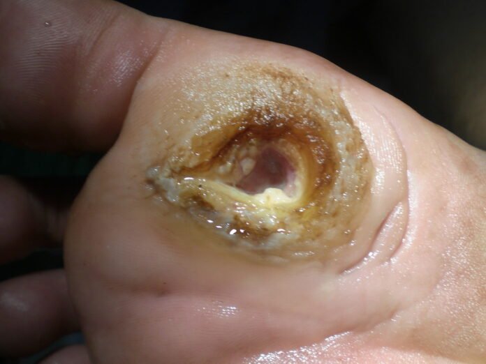
A newly published study funded by the National Institutes of Health (NIH) has revealed a promising diagnostic tool capable of accurately predicting which seemingly healed diabetic foot ulcers (DFUs) are at high risk of reopening. By measuring trans-epidermal water loss (TEWL)—an indicator of skin barrier function—researchers found that wounds with elevated TEWL were substantially more likely to recur. The results, which will be published in the journal Diabetes Care, suggest that incorporating TEWL measurements into standard wound-care protocols could greatly enhance clinicians’ ability to ensure truly complete wound closure and reduce life-long risks associated with DFUs, including lower-limb amputations and premature mortality.
Background on Diabetic Foot Ulcers
Diabetic foot ulcers represent a major and often underappreciated complication of diabetes mellitus. Peripheral neuropathy—a loss of protective sensation in the feet—frequently precludes patients from noticing minor cuts, abrasions, or pressure-induced lesions. Left untreated, these lesions can deepen, become infected, and ultimately lead to tissue necrosis. According to the American Diabetes Association, DFUs account for approximately 85 percent of non-traumatic lower-limb amputations in patients with diabetes. Each year, nearly 200,000 amputations occur in the United States alone due to diabetic foot complications.
Even when a DFU appears to have closed on visual inspection—no open wound, no drainage—underlying tissue layers may remain incompletely healed. This subclinical defect impairs the skin’s barrier function, rendering it unable to retain moisture effectively or block microbial entry. Without a reliable method to confirm true skin barrier restoration, clinicians must rely on visual and palpation exams, which can miss microscopic or deeper-level weaknesses. Consequently, DFUs that seem healed can and often do reopen: recurrence rates within one year of apparent healing range from 30 to 40 percent in various studies. Each recurrence compounds the risk of serious infection, hospital admission, and amputation.
Trans-Epidermal Water Loss as a Predictor
Trans-epidermal water loss (TEWL) quantifies the amount of water vapor passing from the dermis through the epidermis to the surrounding atmosphere. In healthy skin, the stratum corneum (the outermost epidermal layer) acts as a primary barrier that minimizes TEWL. If the skin barrier is compromised or immature—as in a healing wound—TEWL values rise. Historically, TEWL monitoring has been a staple of burn care, where assessing the integrity of newly regenerated skin is critical for treatment planning.
The NIH-funded research team, working under the Diabetic Foot Consortium housed within the NIH’s National Institute of Diabetes, Digestive, and Kidney Diseases (NIDDK), sought to adapt TEWL measurements for evaluating DFUs. As Dr. Teresa Jones, M.D., Program Director in the Division of Diabetes, Endocrinology, & Metabolic Diseases at NIDDK, explained:
“Foot ulcers are such a confounding issue with diabetes, and being able to determine which wounds are at highest risk for recurrence could save many lives and limbs.”
Study Design and Methods
The investigation enrolled more than 400 participants across multiple clinical centers affiliated with the NIDDK Diabetic Foot Consortium. Eligible participants were adults diagnosed with type 1 or type 2 diabetes who had a documented DFU that, by visual standards, appeared to be completely healed (i.e., closed epithelial surface, no drainage). At enrollment—once each DFU was deemed “healed” by routine criteria—researchers recorded baseline data on patient demographics, diabetes duration, comorbidities (e.g., peripheral artery disease, end-stage renal disease), and ulcer history (e.g., ulcer duration, location, prior recurrences).
TEWL Measurement Protocol
Using a standardized, noninvasive TEWL device, clinicians measured evaporative water loss at the site of the healed ulcer on the foot. To ensure consistency, TEWL readings were always taken in a controlled room environment: humidity levels maintained between 40 and 60 percent, ambient temperature between 22 and 24 degrees Celsius, and participants acclimated for at least 15 minutes before measurement. Three consecutive TEWL readings were recorded per wound site, and the average was calculated.
Participants were then followed for 16 weeks post-TEWL measurement. During this period, they returned for biweekly foot exams and TEWL re-evaluations if any sign of breakdown was detected. The primary endpoint was ulcer recurrence, defined as any reopening of the wound at the prior ulcer site, regardless of size.
Key Findings
Among the study cohort, TEWL readings varied widely. Researchers stratified participants into two groups based on TEWL levels:
- High-TEWL Group: TEWL values exceeding 10 grams per square meter per hour (g/m²/h)
- Low-TEWL Group: TEWL values at or below 10 g/m²/h
At 16-week follow-up, 35 percent of participants with high TEWL reported ulcer recurrence, compared to only 17 percent in the low-TEWL category. Statistical analysis revealed that participants in the high-TEWL group were 2.7 times more likely to experience a wound recurrence than those with low TEWL (hazard ratio 2.7; 95% confidence interval 1.8–4.0; p < 0.001).
Further stratification showed that TEWL thresholds could be fine-tuned for greater predictive accuracy. For instance, wounds with TEWL above 12 g/m²/h had a 40 percent recurrence rate, whereas those with TEWL between 8 and 10 g/m²/h exhibited a 20 percent recurrence. Even within the low-TEWL cohort, patients with TEWL values nearing 10 g/m²/h had a slightly elevated risk compared to those below 5 g/m²/h, underscoring a dose–response relationship between TEWL and ulcer reappearance.
Notably, TEWL remained a strong predictor of recurrence after adjusting for confounding variables—including age, glycemic control (HbA1c levels), presence of peripheral arterial disease, and ulcer location—indicating that impaired barrier function was independently associated with wound breakdown.
Clinical Implications
The study’s findings have immediate and far-reaching implications for DFU management:
- Objective Confirmation of Healed Wounds
TEWL measurement offers a quantitative, reproducible method to confirm that a DFU has achieved true barrier restoration—beyond superficial epithelial closure. Incorporating TEWL into routine post-healing assessments could prevent premature discontinuation of protective footwear or offloading devices. - Risk Stratification and Personalized Follow-Up
By identifying patients with higher TEWL—who face a 2.7-fold increased risk of recurrence—clinicians can intensify follow-up regimens. For example, high-TEWL patients might receive more frequent podiatry visits, earlier referral for vascular evaluation, or extended use of wound dressings and pressure-relief insoles. - Targeted Interventions to Restore Barrier Function
Elevated TEWL suggests that underlying dermal layers remain insufficiently healed. Interventions such as prolonged application of advanced wound dressings (e.g., hydrocolloids, silicone sheets), adjunctive topical growth factors, or regenerative therapies (e.g., platelet-rich plasma, skin substitutes) could be tailored to bolster barrier integrity. - Cost-Effectiveness and Health Economics
DFU recurrences impose a devastating financial burden—estimated at $9 to $13 billion annually in the United States for inpatient costs alone. Preventing even a fraction of recurrences through TEWL-guided care could yield significant savings for healthcare systems, insurers, and patients, in addition to reducing morbidity and amputation rates. - Alignment with Burn-Care Practices
In burn units, TEWL measurements guide decisions regarding graft take and timing of topical antimicrobial therapy. Applying similar principles to DFUs aligns diabetic wound care with established burn-care standards, enhancing interdisciplinary knowledge transfer.
Expert Commentary
Dr. Teresa Jones, M.D., Program Director in NIDDK’s Division of Diabetes, Endocrinology, & Metabolic Diseases, lauded the study:
“This study is an important initial step to give clinicians treating diabetic foot ulcers a reliable diagnostic aid for the first time to assess an individual’s risk of ulcer recurrence. Foot ulcers are such a confounding issue with diabetes, and being able to determine which wounds are at highest risk for recurrence could save many lives and limbs.”
Dr. Jones emphasized that diabetes-related amputations carry an 80 percent five-year mortality rate. By reducing ulcer recurrence—and thereby lowering amputation risk—TEWL-based protocols could not only preserve limb function but also extend patient survival.
Pathophysiology of Impaired Barrier Function
Diabetes disrupts normal wound-healing cascades at multiple levels:
- Hyperglycemia-Induced Microvascular Dysfunction
Chronic high blood sugar damages small blood vessels in the dermis and subcutaneous tissues, limiting oxygen and nutrient delivery to healing wounds. Fibroblast proliferation and collagen synthesis become compromised, resulting in thin, fragile scar tissue even after an ulcer appears closed. - Neuropathy and Reduced Innervation
Peripheral neuropathy impairs sensory afferents that normally modulate local blood flow and epidermal turnover. Loss of protective reflexes increases mechanical stress on the lesion, while reduced neurotrophic signaling slows epidermal maturation. - Altered Immune Response
Diabetic patients exhibit impaired neutrophil function, reduced macrophage activation, and defective cytokine signaling. These immune deficits blunt the inflammatory phase of wound healing, delaying progression to proliferative and remodeling phases.
Because TEWL reflects the functional competence of the stratum corneum and underlying lipid matrix, high TEWL in a diabetic ulcer indicates that the new epidermal barrier lacks the structural proteins and lipids that minimize water diffusion. Microtears or immature cornified envelopes allow insensible fluid loss to remain elevated, even if the wound no longer bleeds or exudes serous fluid.
Study Limitations and Future Directions
Despite its robust design and compelling findings, the TEWL study has several limitations:
- Short Follow-Up Period
The 16-week observation window captures early re-openings, but longer-term data (e.g., six months to one year) are needed to determine if TEWL’s predictive power persists over time. - Single-Cohort, Consortium-Based Sample
Although the NIDDK Diabetic Foot Consortium’s multicenter approach enhances generalizability, additional studies in diverse geographic and socioeconomic settings—including rural clinics and underserved urban communities—are required. - Device Standardization
TEWL measurement devices vary among manufacturers. The study used a single, validated instrument. Future research must confirm that different sensors yield comparable results and establish calibration protocols for widespread clinical deployment.
Going forward, the research team plans to:
- Conduct Longitudinal Studies with Extended Follow-Up
Investigators will track healed DFU patients for up to 52 weeks, recording TEWL changes over time and correlating them with ulcer recurrence, amputation incidence, and patient-reported outcomes such as pain and mobility. - Test Interventions to Reduce TEWL
Randomized controlled trials will examine whether targeted barrier-enhancing therapies—such as topical ceramide-based formulations, occlusive dressings, or local photobiomodulation—can lower TEWL in recently healed DFUs and consequently reduce recurrence rates. - Integrate TEWL into Wound-Care Guidelines
Collaborating with professional bodies (e.g., American Diabetes Association, Wound Healing Society), researchers aim to incorporate TEWL thresholds into consensus statements. For example, adding a TEWL < 10 g/m²/h requirement before discontinuing protective offloading or transitioning to standard diabetic footwear. - Develop Mobile TEWL Devices
To facilitate home monitoring, engineering teams are exploring portable TEWL sensors that diabetic patients or caregivers can apply at the bedside. Smartphone connectivity could allow remote clinicians to receive TEWL data in real time, enabling telehealth interventions when barrier function declines.
Diabetic Foot Consortium and Funding Acknowledgments
This landmark study was made possible by grants from NIH/NIDDK (U01DK119099, U24DK122927, U01DK119100, U01DK119083, U01DK119094, U01DK119085, U01DK119102) and carried out by members of the NIH’s Diabetic Foot Consortium. The Consortium brings together multidisciplinary teams of diabetologists, endocrinologists, wound-care specialists, podiatrists, vascular surgeons, and engineers. Its mission is to develop evidence-based innovations that improve the prevention, diagnosis, and treatment of diabetic foot complications.
Conclusion
The identification of TEWL as a reliable predictor of DFU recurrence represents a major advance in diabetic wound care. By revealing subclinical barrier defects in wounds that appear visually healed, TEWL measurements allow clinicians to stratify patients by risk and deploy tailored interventions. Such an approach promises to reduce recurrent ulcer rates, lower amputation incidence, and improve quality of life for millions of diabetics worldwide.
Incorporating TEWL into existing treatment standards can transform the currently reactive model of ulcer management—where recurrences and hospitalizations drive healthcare costs—into a proactive, preventive paradigm. As researchers validate these findings with larger, longitudinal studies and integrate TEWL into clinical guidelines, patients and providers alike can look forward to more secure, durable healing of diabetic foot ulcers. Ultimately, reducing foot ulcer recurrences translates not only to fewer amputations but also to prolonged survival and enhanced mobility for people living with diabetes.
READ MORE: New COVID-19 Variant NB.1.8.1 “Pretty Much Everywhere” in Australia
